Patients who are suspected of having a slipped disc, after a physical examination are asked to undergo an MRI scan. Learn more about this back problem, as well as an MRI scan, right here.
Mild to severe back pain along with leg pain and weakness in the lower extremity muscles is often an indication of a slipped disc. It is a term used by non-medical people, and is actually called spinal disc herniation or herniated disc and is caused due to a tear in the outer ring of an intervertebral disc of the spine, conducing the soft inner part of the disc to bulge out; provoking an inflammatory reaction leading to severe pain. In order to get a proper look at the herniated disc, an MRI scan is conducted.
What is a Slipped Disc
The disc between the vertebrae of the spine is covered in a hard outer shell and contains a jelly-like soft center, which if damaged or ruptured, pushes out the soft inner center into the spinal canal. This reduces the space between the two vertebra and the nerves in the spine, thus, irritating and inflaming the nerves. In some cases, people develop sciatica that leads to lower back pain, buttock pain as well as leg pain, and the pain experienced is continuous. It is usually caused due to aging as with normal wear and tear, the elasticity is lost, making it vulnerable to injury, damage or rupture.
Symptoms
With reduction of space between the nerves, compression is induced, due to which wrong signals are transmitted and lead to symptoms such as:
- Electric shock pain due to compression of the neck or lower back region; the pain is felt down the arms and legs
- Tingling sensation and numbness in the arms and legs
- Weakness of muscles due to interruption of brain signals occurring due to the nerve irritation
- Problems relating to urinating or bowel movements, are generally related to surgeries making it a medical emergency and requiring immediate medical examination
Diagnosis
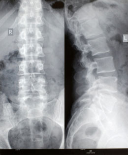
In most cases, it is diagnosed through physical examination. Keeping the medical history in account, lower back, neck, arms and legs and spine movements are inspected; which helps in determination of pain if twisted,bent or folded. The muscles are tested for strength and to determine the source of pain. The tendon reflexes under the kneecap or on the ankle might be tested as well. After the physical examination, diagnostic tests that includes X-ray, MRI and sometimes even a CAT scan are suggested.
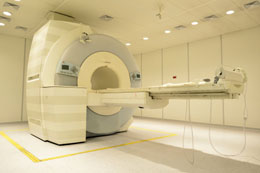
MRI Scan
It is an imaging test and is not suggested until other non-surgical treatments fail to relieve the pain, generally after 4 weeks of treatment. MRI or Magnetic Resonance Imaging does not use ionizing radiation, but radio waves to generate a series of images, depicting thin slices of the body that can be studied from various angles. Thus, any abnormal tissue is better detected and observed.
During the test, the patient is asked to lie down on a table that slides into a huge tunnel-like machine. A contrast material is inserted into the area where the symptoms are observed through an intravenous (IV) line in the hand. Then, the patient is moved into the machine and pictures of the cervical, thoracic and lumbar region are taken; the entire process consumes about 30 minutes to an hour.
MRI scan confirms the location and severity of the slipped disc, and helps in finding details of the spinal infection or presence of a possible tumor. It helps in detecting any tear, herniation or fragmentation of the discs, in detection of spinal stenosis as well as other back problems or any abnormalities of the spine. However, it does not always indicate any serious back or spinal problems. Always speak to your doctor about any doubts you may have when undergoing a diagnostic test.