Decompressive lumbar laminectomy is a surgical procedure used to treat a condition referred to as lateral recess stenosis, which occurs when spinal nerves are pinched by narrowing of the sides of the spinal canal. Symptoms include intense pain as well as numbness and/or weakness in one leg.
This surgical procedure can also be used to treat lumbar canal stenosis, which occurs when the spinal canal becomes narrowed and the cauda equina becomes compressed. This condition has similar symptoms as lateral recess stenosis.
The human spine extends from the skull to the pelvis and is made up of individual bones called vertebrae. The vertebrae, which are stacked on top of each other, are grouped into 4 regions:
The base of the spine (called the coccyx) includes naturally fused vertebrae and is often called the tailbone.
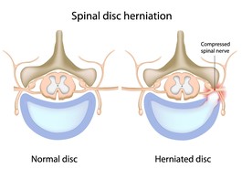
The vertebrae are separated from one another by soft pads, called intervertebral discs, which allow the spine to bend and flex and act as shock absorbers during regular activity. These discs also prevent the vertebrae from rubbing up against each other. Each disc is made up of 2 parts, a soft center called the nucleus and a tough outer band called the annulus.
Throughout the length of the spine is an arch of bone called the spinal canal. Inside the spinal canal are the spinal cord and spinal nerves. The spinal cord begins at the base of the brain and ends in the upper lumbar spine area in a bundle of nerves known as the cauda equina. A pair of spinal nerves branch out (one to the left and one to the right) at each vertebral level. Each nerve exits the spinal canal through an opening between the vertebrae called a foramen.
Healthy discs help to cushion the vertebrae and keep the opening of the foramina wide enough for the spinal nerve roots to pass through without being pinched.
However, as the body ages, the intervertebral discs begin to break down and the vertebrae become much closer to one another. This can result in the formation of bone spurs and cause the spinal canal and the foramina to become narrowed. It also increases the chances that spinal nerves may eventually become pinched. This leads to the need for a decompressive lumbar laminectomy.
The patient is placed under anesthesia and positioned on his or her stomach or side. A small incision is made in the lower back in order for the surgeon to see the pinched spinal nerves and/or the compressed cauda equina.
The surgeon then uses a retractor to expose the vertebrae by spreading apart the muscles and fatty tissue of the spine. A small drill or bone biting instruments are used to remove a section of the vertebra. An opening is cut in the ligamentum flavum in order to reach the spinal canal.
The surgeon removes bone spurs (osteophytes) and any rough edges on the intervertebral disc. This enlarges the foramen and the spinal canal and helps relieve pressure on the spinal nerves.
If necessary, the surgeon will perform a spinal fusion with instrumentation to help stabilize the spine. A spinal fusion involves grafting a small piece of bone (usually taken from the patient's own hip) onto the spine and using spinal hardware, such as screws, rods, or other metal implants, to support the spine and provide stability.
Then the procedure is finished! The surgeon will close the incision either using absorbable sutures (stitches), which absorb on their own and do not need to be removed, or skin sutures, which will have to be removed by the surgeon after the incision has healed.
Copyright © www.orthopaedics.win Bone Health All Rights Reserved