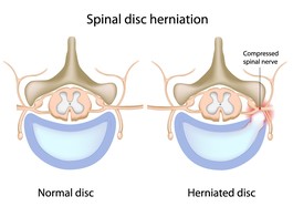
Cervical stenosis means, literally, tightening or narrowing of the canal around the spinal cord. Of the degenerative disorders that can affect the spine, it is potentially the most serious. If the cervical stenosis is profound enough, it can cause dysfunction of the spinal cord known as myelopathy. The typical person who has cervical stenosis and myelopathy may be in his or her fifties or early sixties. The patient often has complained of neck pain for many years. In some cases, the pain can actually be mild. Therapy may have been prescribed, in addition to medications, for the pain. The other features of this cervical spine disorder will be demonstrated in an illustrative case.
Mrs. S is a 61-year-old woman with a long-term complaint of neck pain. In the past, her pain has been amply controlled with ibuprofen (Motrin) and some home exercises. Occasionally, she wears a soft cervical collar to calm her neck spasms. She is an avid knitter and has made several sweaters and scarves for her grandchildren recently. In the past two months, however, she finds that her fingers are becoming clumsy, and she has to take frequent breaks. In addition, Mrs. S is finding that she is not as agile buttoning her shirts in the morning. She is not complaining of any pain in the arms or legs. Interestingly, her legs are a bit wobbly, but she attributes that to some arthritis that has set in over the years. Mrs. S has had no problems urinating on her own, and no change in her bowel habits or control.
Examination
At the doctor's office, she is given a full examination. She has a somewhat decreased range of motion of the neck, with some pain at the extremes of the movements. She walks with an abnormal gait, which can be described as "wide-based." In looking at her feet during ambulation, her feet are more spread apart than normal and she stumbles a bit with some steps.
Her reflexes in her arms and legs are very jumpy. This is termed hyperreflexia. She does not have any noticeable weakness in the arms or legs. Because of these findings, the doctor gets some x-rays in the office.
Diagnostic Tests
The plain x-rays show a degenerative spine. This can be better termed as spondylosis. She has osteophytes in the front and back of the spine, which might be protruding into the spinal canal. From the x-ray, there does not appear to be any masses or lesions that would suggest a tumor or infection. Mrs. S's doctor knows that these bony changes are very common at her age. Also, he understands that the plain x-ray is not a very good way of assessing the spinal cord or the space around it since it only shows the bony structures, not the soft tissue structures.
Mrs. S is sent for an MRI scan of her neck. This test entails her lying down for about 45 minutes in a long-tube. The long-tube has a very large magnet in it. This is what is responsible for the magnetic part of the magnetic resonance imaging (MRI). Because different tissues in the body respond to magnetic fields in different ways, they have characteristic appearances on MR images.
Mrs. S's MRI showed severe narrowing of her spinal canal. Most of this narrowing is coming from degenerated discs that are protruding into the spinal canal. These discs appear hard and have bony osteophytes above and below them, making the compression even less forgiving.
Specialist Consultation
After getting the MRI report, Mrs. S's doctor sends her to a spine surgeon. She explains to the patient that her condition is called cervical spinal stenosis. Because her stenosis, or tightening, is severe, the nerves in the spinal cord cannot function normally. The compression of the spinal cord is causing her to fumble with her knitting needles and shirt buttons, as well as giving her "wobbly" legs. This surprised Mrs. S the most, as she was sure that she had knee arthritis that was causing her leg symptoms. The spine surgeon explained that the nerves that go to both the arms and legs pass through the neck within the spinal cord. Thus, compression at the neck can cause symptoms in the arms and legs.
Mrs. S asks what can be done about her condition, and the spine surgeon explains that it is likely that her finger and leg fumbling can get worse. In fact, the tightening around the spinal cord can get to a point that she may lose control of her bladder and bowels. In the best case scenario, her symptoms will stay the same for the rest of her life, which can be expected in a low percentage of patients.
The treatment options given to Mrs. S are that she can be treated non-operatively or by surgery. The surgeon explains what comprises non-operative treatment. It includes nonsteroidal medications (like Motrin, Naprosyn, Celebrex, or Vioxx), physical therapy for the neck muscles, cervical collar use, and traction. Of the options, Mrs. S was most concerned about traction, as she would have to be lying down for a portion of the day while the weights were attached to her chin and head.
Mrs. S was also informed of the surgical options. Because of the extent of her disease, the surgeon explained that the best method of relieving the pressure on the spinal cord was to remove the bone from the front of the neck and off the spinal cord. This is known as a corpectomy (core-peck-toe-me). This would entail an incision in the front the neck through which the surgeon can remove the parts of the vertebral bodies that are compressing her spinal cord. In place of the vertebral bodies, a large piece of bone from her own pelvis, or a cadaver donor, would be inserted. This bone would be expected to heal in place. This is known as a fusion. The likelihood of catching any disease from the cadaver bone is extremely low and is in fact much lower than contracting any disease from a blood transfusion. The more significant risks were from the surgery itself, she was told. The possible complications include damage to the large arteries that supply blood to the brain and to the spinal cord. Spinal cord damage may cause Mrs. S to be completely paralyzed from the neck down. These are the most serious complications; she was informed.
Other possibilities, like infection are also possible, but are more easily treated. Damage to the nerves that supply the vocal cord is also a potential complication. Mrs. S was made aware of this possibility, and that she could have hoarseness permanently after the operation. After hearing the options, Mrs. S asks the spine surgeon a few key questions. First, if she has damage to her spinal cord already, what are the chances of her symptoms getting better with surgery?
Because she is still highly functional, she has a good chance of resolving some, though perhaps not all, of her neurologic symptoms. Her neck pain, though not the focus of the surgery, may or may not get better. If the surgery is a complete success, she will be able to return to her previous activities with a greatly decreased chance of her spinal cord being compressed further. In essence, the surgery is mostly to keep her from progressively getting worse and/or prevent a catastrophic event like a spinal cord injury, which could result with a very minor injury such as a fall or slip.
What will happen to her if she doesn't choose surgery? From the studies available, it is probable that Mrs. S's cervical stenosis will worsen with time. Although it is possible that she could live the rest of her life without any advancement of her problems, it is unlikely. Furthermore, it is even more unlikely for her neurologic symptoms to significantly improve.
What happens if the piece of bone doesn't heal in place? This is a rather common complication, occurring in about 15 to 20 percent of patients undergoing this procedure. In the event that the bone doesn't fuse in the front, a second surgery to fuse the back of the vertebra is performed. This is done by an incision along the back of the neck.
Outcome in Cervical Spinal Stenosis Case
Weighing the options and contemplating the possible complications of both operative and non-operative treatment, Mrs. S decides to proceed with surgery. Thankfully, the surgery was without complication. After surgery, she remained in the hospital for three days. Her neck was very sore, but strong pain medication helped manage the pain.
She was instructed to keep a hard cervical collar on at all times for six weeks. She was able to get out of bed the day after surgery and started eating a full diet, as she was able to tolerate. After she was discharged, she followed up with her surgeon in the office. The wound healed well. After six weeks she did not use the cervical collar anymore. The bone graft showed good signs of healing to her own bone on the x-rays. After three months, she felt that her fingers were working better and she no longer felt wobbly in the legs. She returned to knitting, producing a blue baby bonnet for her newborn grandson.
Garfin and Bono have provided an eloquent dissertation for the consumer regarding degenerative cervical spine disorders. They have provided a discussion that encompasses the myriad of diagnoses and potential problems afflicting the degenerating and aging cervical spine.
They have outlined the anatomy and covered the majority of pathological processes that afflict the cervical spine. For the symptomatic consumer, the information is particularly revealing in that they provide insightful "first hand information" regarding the decision-making process that requires the active participation of both the patient and physician.
This is a "must read" for patients considering surgical intervention for cervical degenerative disease. For providing this information, Drs. Garfin and Bono are to be heartily congratulated.
Copyright © www.orthopaedics.win Bone Health All Rights Reserved