Diagnosing spinal stenosis can be challenging. The symptoms can mimic those of other conditions, plus the symptoms can come and go. To figure out the cause of your spinal stenosis, your doctor will need to perform several exams and tests. These exams and tests will also help the doctor develop a treatment plan for you—a way to manage your pain and other symptoms and to help you recover.
During your visit, your doctor will ask about your current symptoms and remedies you have already tried. This is part of your physical exam. He or she will ask some typical questions, such as:
You may also need to have some imaging tests done to help your doctor diagnose the cause of your spinal stenosis. These diagnostic studies are usually performed if symptoms do not subside after a period of 3 to 6 months of therapy such as rest, anti-inflammatory medications, and physical therapy.
Imaging tests are ordered cautiously because many people who do not have any symptoms of spinal stenosis have abnormal x-rays, CT scans, and MRIs. Surgery should only be performed in patients whose symptoms correlate with findings on these studies and a history that supports these findings.
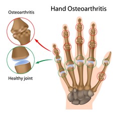
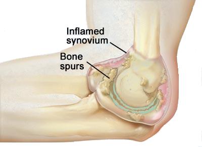
Typically, plain x-rays are done first. They are helpful in looking for infection, tumors, and identifying problems with alignment of the spine. They can show narrowed disc space, fractures, bone spurs (osteophytes), or osteoarthritis (spondylosis).
A computerized axial tomography scan (a CT or CAT scan) or a magnetic resonance imaging test (an MRI) can show a bulging disc or a herniated disc. An MRI image is shown below.
You also may be asked to undergo additional tests, such as:
All of this information—the physical exam and the imaging tests—will help your doctor plan how best to treat your spinal stenosis.
Copyright © www.orthopaedics.win Bone Health All Rights Reserved