Today new technological advancements in thoracoscopy have made it possible for thoracic scoliotic curves to be corrected utilizing minimally invasive techniques. Thoracoscopy combines the science of endoscopy (video-assisted surgery) with thoracotomy (access to chest, or thoracic spine).
Utilizing thoracoscopy, highly skilled spinal surgeons have determined lateral (from the side) entry through the chest wall provides sufficient access to the thoracic spine to enable thoracic scoliotic curve correction.
Traditionally, surgical treatment for thoracic scoliosis meant an open procedure leaving the patient with a large unsightly scar. Instead of an open procedure (large incision), the spine surgeon makes small incisions - precisely located - to allow access to the thoracic spine. Tiny, specially designed endoscopic instruments pass through these incisional gateways and are maneuvered during surgery.
This is exciting news for patients with progressive scoliotic curvature because minimally invasive procedures offer so many benefits to the patient. During an open procedure, the incisions slice through skin, layers of fat, and muscle, which are then held back by clamps or pulled aside with retracting instruments. Blood vessels are sealed to prevent serious loss of blood. Conversely, patients who undergo a thoracoscopic procedure find:
The spine surgeon selects the procedure(s) that will provide the most benefit to the patient. Scoliotic curve reduction usually involves the removal of several intervertebral discs (discectomy), spinal instrumentation, and fusion.
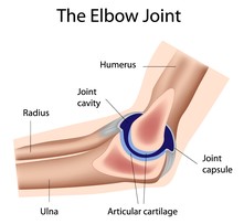
Spinal Instrumentation and Fusion are surgical procedures utilized to correct spinal deformity and to provide permanent stability to the spinal column. These procedures join and solidify the level where a spinal element has been damaged or removed (e.g. intervertebral disc). Instrumentation utilizes medically designed implants such as rods, wires, and screws. These devices hold the spine in place during fusion. Fusion is the adhesive process joining bony spinal elements.
The patient's history is reviewed and a physical examination is performed. The patient's pelvis and shoulders are observed for differences in height, and spinal rotational flexibility. Posterior/anterior (P/A) and lateral x-rays are obtained along with side bending films. The degree of curvature is measured and marked utilizing a full-length A/P x-ray.
General anesthesia is administered to all patients. The anesthesiologist decides the type of anesthesia based on many variables including the patient's age and weight. The patient is positioned on the operating table with the curvature side up (concave side down). The hips and shoulders are taped into place securing the patient in the proper position for surgery. The patient's position is checked throughout the procedure. A C-Arm is a movable fluoroscopic unit that projects a selected view of the spine onto a monitor located at the foot of the operating table. As the C-Arm is moved around the patient, the surgeon gains information important to determining spinal landmarks (guides), as well as spinal rotation. The landmarks represent specific areas where incisions will create a portal. A portal is an opening through which tiny specialized surgical instruments are inserted. These entry points (landmarks) are marked directly onto the patient's skin.
C-Arm Use Landmarking Patient's Skin Landmarks or PortalsLandmarking Patient's Skin
Landmarks or portals are created for precise entry into the chest. An endoscopic camera sends images to two monitors utilized during the surgery. Each monitor is placed near the patient's head, one anterior and the other posterior. This allows the surgeon and his or her assistant to view the procedure.
Surgical Procedure
The portal incisions are made, an endoscopic camera is inserted into the chest, and surgery begins as tiny specialized instruments are maneuvered through small hollow tubes. According to the surgical plan, several intervertebral discs and adjoining endplates are removed using a procedure termed discectomy.
Typical Monitor Set-up in Operating RoomDiscectomy
Intervertebral Disc Removal
Endplate Removal
After the removal of necessary discs and endplates, the empty space is examined using a scope and later packed with bone graft harvested from the ribs. Bone graft is necessary for fusion. Discectomy is followed by spinal instrumentation. The C-Arm is the surgeon's anatomical guide assisting with many measurements associated with screw and rod placement.
DiscectomyC-Arm Fluoroscopic View of Vertebral Screw Insertion
After the rod has been cut to the correct length and affixed to the screws, the spine is compressed. A rack and pinion device fits over two screw heads on the rod and is turned distributing compressive force evenly to correct spinal alignment. When the vertebral alignment is corrected, the screws are plugged to hold the rod in proper position. The endoscopic instruments and camera are removed and the small incisions are closed and covered with small dressings. Before the patient is transferred to the recovery room, A/P and lateral x-rays are taken to record instrumentation and curve correction.
C-Arm Fluoroscopic View of Vertebral Screw InsertionThoracoscopic correction of thoracic scoliosis is a remarkable advancement in corrective spinal surgery. Most patients are out of bed walking the day following surgery and released from the hospital within a few days. Although patients may be braced for 3 months, recovery is rapid enabling most patients to return to many regular activities. Children often return to school within 2 to 4 weeks. Patient progress is monitored during one, three, six, and 12-month intervals, which include an x-ray evaluation of the spine.
Copyright © www.orthopaedics.win Bone Health All Rights Reserved