Your back, or spine, is made up of many parts. Your backbone, also called your vertebral column, provides support and protection. It consists of 33 vertebrae (bones). There are discs between each of the vertebra that act like pads or shock absorbers. Each disc is made up of a tire-like outer band called the annulus fibrosus and a gel-like inner substance called the nucleus pulposus. Together, the vertebrae and the discs provide a protective tunnel (the spinal canal) to house the spinal cord and spinal nerves. These nerves run down the center of the vertebrae and exit to various parts of the body.
Your back also has muscles, ligaments, tendons, and blood vessels. Muscles are strands of tissues that act as the source of power for movement. Ligaments are the strong, flexible bands of fibrous tissue that link the bones together, and tendons connect muscles to bones and discs. Blood vessels provide nourishment. These parts all work together to help you move about.
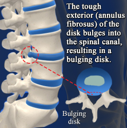
A herniated disc most often occurs in the lumbar region (low back). This is because the lumbar spine carries most of the body's weight. Sometimes the herniation can press on a nerve, causing pain that spreads or radiates to other parts of the body. The amount of pain associated with a disc rupture often depends on the amount of material that breaks through the annulus fibrosus and whether it compresses a nerve.
Copyright © www.orthopaedics.win Bone Health All Rights Reserved