Calcaneal stress fracture is one of the most common problems associated with the tarsal bone. The common symptoms are inflammation in the heel, inability to bear weight on the heel, and bruising of the affected area. The treatment may include rest, partial to complete immobility, application of plaster, and surgical procedures.
Calcaneal bone refers to the bigger heel bone which is located at the back of the foot. The main function of this bone is to maintain the vertical posture and enable one to walk properly. It acts as a foundation and supports the total weight of the individual. It also supports the lateral column of the foot. The outer layer of the bone comprises a tough cortical bone that encloses a soft cancellous bone inside. This specific structure makes the calcaneus bone prone to injuries. An overuse of this bone may lead to calcaneal stress fractures.
Causes and Symptoms
These fractures are common among people who participate in activities that involve recurrent use of the heel bone, such as marching, jumping, climbing, and dancing. Hence, military people, dancers, rock climbers, roofers, and other athletes are at a higher risk of these fractures. A fall from a height of about 6 feet or more often results in this fracture. Another common cause of this condition is sudden injury and/or accidents.
The severity of the fracture depends upon three factors, namely, the position of the bone during the fracture, the orientation of the force that caused the fracture, and the strength of the bone. Based on the severity of swelling within the bones, the fracture is categorized under various groups. Some of the noticeable symptoms of this condition include swelling and inflammation in the heel area, inability to put weight on the heel, and at times bruising of the bone and the surrounding portion. Usually, heel pain worsens on walking and lifting weights, and continues to remain for a long period.
Diagnosis and Treatment
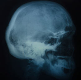
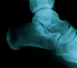
At times, these fractures remain undiagnosed even though there is characteristic heel pain. Initial radiographic results of the heel portion often show normal structure. However, diagnosis can be confirmed by conducting repeated radiographic examinations after every 4 weeks. The orthopedist may also conduct imaging tests such as X-ray, MRI (magnetic resonance imaging), and CT (computed tomography) scans from different angles in order to identify the severity of the fracture.
These fractures are more difficult to treat compared to other tarsal bone fractures. In fact, it is one of the most challenging cases in orthopedics. However, if left untreated, it may result in deformity of the gait. If an individual is diagnosed with this fracture, the most effective pain management and healing procedure is to rest for at least 6-8 weeks. During this period, the patient may use crutches for standing, walking, and other locomotive activities. Other treatment and/or management options of these fractures may vary depending upon the severity and the type of fracture.
In case of a severe undisplaced calcaneal stress fracture, the orthopedist may prescribe application of a plaster cast in order to keep the heel bones in place. Such a treatment method should be continued until radiographic results show improvements in the bone healing process. For displaced calcaneal stress fractures, closed reduction to manipulate the fractured bone may be considered to treat the condition. If the patient is not responsive to this method, the physician may conduct a surgical procedure and/or an open reduction.
Disclaimer:
This Buzzle article is for informative purposes only, and should not be used as a replacement for expert medical advice.