Question
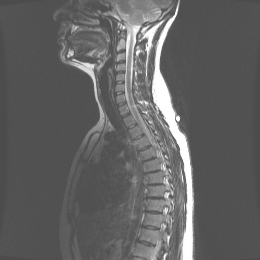 MRI
MRI 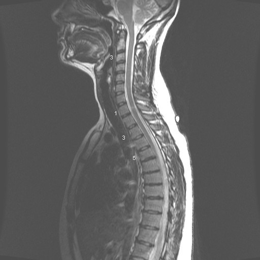 MRI 2
MRI 2
Hello and thank you for taking your time to read this and I apologize for the length of this. :) I am a 30 year old female and I have been having chronic back pain for quite a few years with the pain increasing in intensity steadily for the past 3 years. I used to figure skate competitively from age 10 to 17. While I did not sustain any major back injuries during that period, I think it would be safe to say my spine definitely did things it was never designed to do for long periods of time. I have seen a few doctors regarding this matter, one of which said something to the effect of "i don't see anything unusual here" regarding my MRI results while another's opinion was "you are too young to have these problems in your spine" and a nurse in the office of the MRI ordering Dr. who said "I don't know how you even move". I've tried to decipher the MRI reading myself but have trouble putting it all together.
------------------------------------------------
The summary of my MRI as follows (age 27 at time of MRI):
THORACIC SPINE MRI WITHOUT IV CONTRAST, 9/24/2010 CLINICAL HISTORY: Chronic mid back pain. Multiple injuries. Bilateral lower extremity pain and numbness. COMPARISON: None. FINDINGS: There is a mild thoracic scoliosis that is convex right and centered at the T7-T8 level. The thoracic vertebral body heights and anterior-posterior alignment appear unremarkable, without evidence of compression fracture or spondylolisthesis. There are multilevel anterior marginal osteophyte formations most notably in the midthoracic spine between T6 and T9. There are mild multilevel facet joint degenerative changes in the thoracic spine. There is a small left lateral T5-T6 disc protrusion that results in mild central spinal canal stenosis. There are tiny right paracentral T6-T7 and T7-T8 disc protrusions. There is a tiny left paracentral T8-T9 disc protrusion. There is a small right lateral T11-T12 disc protrusion that results in mild right lateral recess stenosis. There is mild multilevel disc bulging in the thoracic spine. There is no significant neural foraminal stenosis in the thoracic spine. The thoracic spinal cord appears unremarkable in size and signal intensity, without evidence of cord compression or edema. The conus medullaris terminates at the L1 level and appears unremarkable in size and signal intensity.
IMPRESSION: Small left lateral T5-T6 disc protrusion, which results in mild central spinal canal stenosis. Tiny right paracentral T6-T7 and T7-T8 disc protrusions. Tiny left paracentral T8-T9 disc protrusion. Small right lateral T11-T12 disc protrusion, which results in mild right lateral recess stenosis. Mild multilevel disc bulging and facet joint degenerative changes in the thoracic spine. No significant neural foraminal stenosis in the thoracic spine.
FINDINGS: Examination of the alignment of the lumbar spine demonstrates it to be unremarkable. Bone marrow signal, spinal cord signal, nerve roots, posterior elements and surrounding soft tissues are within normal limits. A hemangioma of the left posterior superior corner of the L4 vertebral body is evident and of no clinical concern. T11-T12: A small right central disc protrusion is present spanning approximately 9 mm along the margin of the disc, protruding posteriorly by approximately 3 mm best seen on axial T2 image 4. This minimally impinges upon the ventral aspect of the thecal sac, but does not cause significant central canal narrowing. Facet joints appear unremarkable. There is no significant foraminal narrowing. Disc protrusion probably minimally contacts traversing nerve roots. T12-L1: The central canal and foramina are unremarkable. L1-L2: The central canal and foramina are unremarkable. L2-L3: The central canal and foramina are unremarkable. L3-L4: No significant disc bulge is present. Mild facet arthropathy and trace left facet joint fluid is evident. There is no significant central canal or foraminal narrowing. L4-L5: A small central disc protrusion is visualized. High-intensity zone associated with the posterior margin of this protrusion is evident likely indicative of a small annular tear. Mild facet arthropathy and ligamentum flavum hypertrophy are evident. There is mild central canal narrowing, and minimal bilateral foraminal narrowing. Protrusion minimally contacts but does not significantly displace the traversing right L5 nerve root (axial T2 images 12). L5-S1: No significant disc bulge is present. Facet joints do not demonstrate degenerative change, but STIR images demonstrate trace fluid within the facet joints. IMPRESSION: 1. Small right central T11-T12 protrusion and small central L4-L5 protrusion. L4-L5 protrusion minimally contacts traversing right L5 nerve root. 2. Mild facet arthropathy and trace facet joint fluid of the lower lumbar spine.
------------------------------------------------------------
I was "prescribed" marijuana in 2011 for medical use and that seemed to relieve a lot of the pain but had to stop using it as I had become pregnant and stopped using all drugs for obvious reasons. I am now taking at least 400 mg of ibuprofen daily to take a bit of the edge off. I have tow very active young children and need to be constantly moving, lifting, etc. and it's not fun when your back is searing with pain. I'm not sure of what course of action to take in this matter as I haven't seen a doctor who thought my pain was worthy of any pain medication other than something OTC which I've tried everything under the sun in those regards. I have also seen a chiropractor for weekly adjustments for 4 weeks and follow up monthly visits for 5-6 months thereafter. I've used heat, ice, stretching, exercise, and massage therapy with no substantial or lasting improvement. I'm getting to my wit's end with the chronic pain and I just don't know what to do anymore. The doctors I've seen thus far have been most unhelpful in the matter.
Thanks again for your time, it is immensely appreciated!
AnswerHi Sarah,
Wow, sorry to hear about your pain issues. The differing opinions by the doctors and nurses, while frustrating, is not really unusual. Reading an MRI is a bit of art, opinion, and science so there can be a wide range of opinion.
The good news is I can see why each doctor said what they said, and I do think there is probably a lot that can be done to help you.
People with chronic pain are often in a place where "nothing is wrong," but there is also a whole lot that is "not right." There is nothing "wrong" in the sense that you aren't riddled with bone spurs or have spinal stenosis, BUT there is a whole lot that is "not right" with minor disc bulges and compressions, etc. throughout your spine.
So that you understand where I will be pointing you, take a moment and think of all the muscles in your body like the rigging (ropes) on a big old sailboat. It is the interplay of the tension in our muscles that allows us to stand, sit, walk, pick up a glass, etc. BUT just like the rigging on a sailboat, if one of the pieces of rigging is set too tight it can pull the masts (bones) out of position. And if many of the pieces of rigging are too tight from extreme use or injury in the past then your vertebrae can be pulled and yanked into a myriad of inappropriate and painful positions.
Once you understand the concept that "bones only go where muscles pull or hold them" (bones are merely chunks of calcium that do not move on their own), then you can see that releasing the inappropriate tension throughout your body, but especially in your back, will allow your vertebrae the appropriate space and freedom they require for your nerves and discs not to be compromised and cause your pain.
Okay, so I'm going to recommend two things. You can do either one, or both of these things and I do think that both of them will help you. First, I'm going to recommend that you go to my totally free website,
http://www.do-it-yourself-joint-pain-relief.com/
and follow along with the videos so you can begin to learn how to fix yourself. You may want to start with the Lower Back Pain page(s) when you begin,
http://www.do-it-yourself-joint-pain-relief.com/lower-back-pain-relief.html, since getting your lower back free will help other things let go as well. And feel free to check out the Site Map page where I list every single page on the website,
http://www.do-it-yourself-joint-pain-relief.com/joint-pain-relief-site-map.html
This is a great place to start.
Second, I'm going to recommend you find a Structural Integration practitioner and have them work on you. I'd do my best to find someone who has been doing it at least 5 years, and sounds like they know what they are talking about on the phone, and not to be sexist, but you may want to find a male (or at least a strong woman) if you have the choice, since sometimes strength can be an issue.
The practitioner may want to do your whole body first, but tell them you need a lot of "sixth hour" work first for a least a few sessions. I do think that this work could help you tremendously.
To find a practitioner, go to these two websites:
http://www.rolf.org/find/byLocation?city=&state=WI&country=USA&name=
and
http://www.rolfguild.org/cgi-bin/DB/search.cgi?setup_file=practitioners.setup&se
Call a bunch of these people, and feel free to try more than one if you aren't getting results from your first choice. Like reading an MRI, Structural Integration is both an art and a science, so it matters a great deal who is doing the work.
I do sincerely hope this information helps.
All the best,
Gary Crowley