Streeter dysplasia is a term that historically has been used to describe a complex disorder characterized by constricting rings, acrosyndactyly, or, often, amputations of the extremities of neonates. It is analogous to constriction band syndrome or amniotic band syndrome (ABS), which was recognized as early as 300 BC.
Depending on the severity of the constriction, the defect may be as minimal as a merely cosmetic band. Deeper bands may cause lymphatic obstruction leading to edema and vascular compromise that necessitates immediate release. Pressure from the bands may potentially cause abnormalities distal to the constriction, such as hemihypertrophy, anterolateral bowing, pseudarthrosis, leg-length discrepancy, and resistant teratologic clubfeet. These conditions may lead to limited function and difficulty with ambulation.[1]
Constriction bands across the head and face may lead to facial clefts. If the cleft extends into the cranium, encephaloceles may result. Bands that cross the body may compromise the chest (thoracoschisis or extrathoracic heart) or abdomen (gastroschisis).
Hippocrates suggested that extrinsic pressures from a ruptured amniotic membrane lead to the formation of bands or digital amputations. In 1652, van Helmont reported on intrauterine amputations, which he attributed to the pregnant mothers having looked upon maimed soldiers. Montgomery[2] in 1832 and Simpson[3] in 1836 subsequently described series of amniotic band–associated deformities and discussed the differences between agenesis- and amniotic band–induced amputations.
The condition was not referred to as Streeter dysplasia until 1930, when Streeter postulated a germ plasm defect as one plausible etiology.[4] At that time, this theory was well accepted because of the associated anomalies, which occurred far from the site of the constriction bands. In 1960, Patterson used histology to show how constriction bands look like normal skin creases. He hypothesized that the same lack of mesodermal development occurs in the area of the band, thereby making the bands simply abnormal creases.[5]
Patterson's theory was later refuted by Torpin, who examined many placentae and infants with the disorder.[6] In 1965, Torpin reintroduced the idea originally held by Hippocrates. He proposed that maternal trauma leads to rupture of the amniotic membrane, which then forms into strands. These encircling strands cause extrinsic compression on the head or limb, leading to the formation of bands, vascular occlusion, and, eventually, amputations. Currently, this is the most widely supported hypothesis; therefore, ABS would be a more accurate name for this disorder than Streeter dysplasia.
NextThe anatomy relevant to surgery for ABS depends on the area of the body affected. Most bands are superficial, and only the skin and subcutaneous tissue are involved. However, the neurovascular bundle may be displaced near areas of banding. Therefore, this possibility miust be taken into consideration with dissection for excision in order to avoid devascularization or denervation of the involved body segments.
The developing embryo sits within two cavities, the amnion and the chorion. As development occurs, the amnion presses against the extracoelomic space, eventually obliterating it and bringing the amnion up to and supported by the chorion. This phenomenon occurs on or about week 12 of gestation. Incomplete obliteration of the extracoelomic space renders the amnion fragile and subject to spontaneous or traumatic rupture.[7] After the rupture, a transient oligohydramnios occurs due to extravasation of amniotic fluid. Until the chorion adjusts to the permeability, the developing fetus has very little room in which to move. This may contribute to the severity of clubfeet deformities seen with ABS.
This decrease in space also allows the resultant floating amniotic bands to easily ensnare a developing body part. Early in gestation, the encircling bands may result in spontaneous abortions. If the constriction occurs after development is nearly complete, only fissures, acrosyndactylization, andr intrauterine amputation are noted on the extremities as typical manifestations. If the amniotic bands are swallowed while still partially attached to the placenta, the tether may lead to bizarre facial clefts and palate deficiencies.[8]
Two main lines of thought exist regarding the etiology of amniotic band syndrome, attributing the condition either to intrinsic or to extrinsic causes. Streeter proposed that a disruptive event occurs during blastogenesis, leading to an intrinsic germ plasm defect.[4] This causes the soft tissue to slough. External healing of the slough leads to the constricting rings and the resultant localized developmental defects. Cases of ABS in which the amnion is intact support this theory, as do the frequently associated renal abnormalities (37% of cases) and occasional cardiac abnormalities.
Ainhum, a predominantly African syndrome in which progressive circumferential ulceration leads to eventual amputation of digits, is an inherited disease, thus further lending support to an intrinsic developmental defect as a cause of digit amputation. Streeter aggressively defended this theory for more than 35 years. For his long and avid support, many surgeons still describe this disorder as Streeter dysplasia.
The lack of family history or predictable recurrences in families of children born with ABS negates the theory of an inherent or genetic component to the condition. Zionts et al[9] were able to demonstrate variable findings of the syndrome in monozygotic twins who each were affected by amniotic bands. These and other sporadic findings are more consistent with the current thought that external compression is the etiologic root of this syndrome.[10]
In 1965, Torpin reintroduced the extrinsic theory for ABS.[6] In his study of fetuses and placentae, he noted the lack of a complete amniotic lining in the placentae of neonates with ABS. Strands of amnion were also visible around constricting rings of the digits, and binding strands were visible at the tips of limbs with acrosyndactyly. He proposed that intrauterine trauma led to premature rupture of the membranes, and strands of residual membranes could encircle the digits or might even be swallowed. The presence of severed but otherwise fully developed limbs in newborn infants further validates this extrinsic band compression theory.
A sonographic study by Barzilay et al suggested that prior uterine surgery may be a risk factor for ABS.[11]
The estimated frequency of ABS in the United States is 1 per 3000 pregnancies (if the potential miscarriages that occur from severe banding early in gestation are considered). More common statistics show an incidence of 1 case per 10,000-15,000 population. One of the few epidemiologic studies is a 1988 study from Atlanta that cites the incidence as 1.16 cases per 10,000 population. No sex predilection is recognized.
The incidence overseas is similar to that in the United States. The birth defect registry of Western Australia cites an incidence of 1.15 per 10,000 population.
The prognosis for patients with isolated superficial extremity bands is good (see the image below). Aside from cosmetic variability, no functional deficits remain.
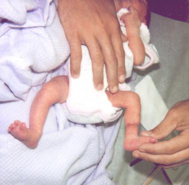 Image shows the lower limb of a young child born with moderate bands that extend deep to the fascia but do not compromise the neurovascular system.
Deeper bands may be associated with progressive problems leading to later lymphatic and neurovascular compromise that necessitates surgical intervention. For patients with acrosyndactyly, hand function is limited secondary to stiffness of the joints, but good prehension and grasp may be obtained with reconstructive procedures. Children who have had intrauterine amputations are usually well adapted to their physical limitations, and often little needs to be done. In children with a transverse deficiency proximal to the ankle joint, a prosthesis is required for full function.
Clinical Presentation
Copyright © www.orthopaedics.win Bone Health All Rights Reserved