Polydactyly is the most common congenital anomaly of the forefoot.[1, 2, 3, 4, 5, 6, 7] It most commonly refers to the presence of six toes on one foot, but more toes are possible.
Polydactyly may be associated with syndactyly. It most frequently occurs as an isolated trait with autosomal dominant inheritance and variable penetrance. Other patterns of inheritance,[8] sporadic occurrence, and association with syndromes are also possible.[1, 9, 10]
Accurate understanding of the involved anatomy leads to better clinical results. Radiographs obtained after ossification of the involved bones allow for definitive treatment of the duplicated parts and all associated abnormalities. Greater appreciation of the longitudinal bracket epiphysis, often seen in the first metatarsal in children with preaxial polydactyly, and descriptions that allow for earlier diagnosis and correction of this abnormality lead to better outcomes.
Treatment for polydactyly of the foot has changed little over time. Removing the extra digit by disarticulation is the standard treatment.[3, 11, 12, 13]
NextPolydactyly may be divided into three broad categories as follows:
Postaxial polydactyly is the most common form,[6] occurring in 80% of cases, followed by preaxial polydactyly (see the image below) and then central polydactyly. The duplication may range from a well-formed articulated digit to a rudimentary digit. Abnormalities of the associated metatarsal commonly occur in polydactyly.
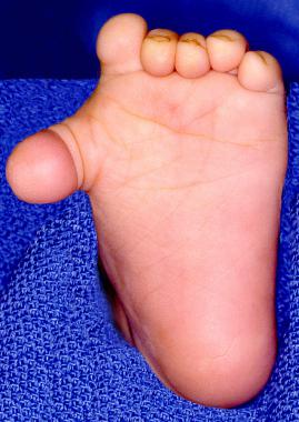 Preoperative photograph of a 1-year-old child with preaxial polydactyly and significant varus of the duplicated toe.
Other, more detailed classification systems have also been described (see Presentation).
Polydactyly may occur as an isolated trait or in conjunction with certain syndromes, and there is a positive family history in 30% of cases. The syndromes with which polydactyly has been associated include Ellis-van Creveld syndrome,[14, 15] trisomy 13, tibial hemimelia, and trisomy 21.
Adam et al observed clinical findings in 18 cases of diabetic embryopathy and preaxial hallucal polydactyly to identify the features most suggestive of diabetic embryopathy.[16] The authors found that proximally placed preaxial hallucal polydactyly, particularly when coupled with segmentation anomalies of the spine and tibial hemimelia, is highly suggestive of diabetic embryopathy. They added that diabetes in the mothers pointed to a possible genetic predisposition interacting with teratogenic effects of poor glycemic control.[17]
The incidence of polydactyly is 1.7 cases per 1000 live births. The frequency is higher in blacks (3.6-13.9 cases per 1000 live births) than in whites (0.3-1.3 cases per 1000 live births). Polydactyly is bilateral in 50% of cases and has a slight male predilection.
Clinical Presentation
Copyright © www.orthopaedics.win Bone Health All Rights Reserved