Madelung deformity (MD) of the wrist is characterized by a growth disturbance in the volar-ulnar distal radial physis that results in a volar and ulnar tilted distal radial articular surface, volar translation of the hand and wrist, and a dorsally prominent distal ulna. It occurs predominantly in adolescent females who present with pain, decreased range of motion, and deformity. It often has a genetic etiology and is associated with mesomelic dwarfism and a mutation on the X chromosome. The deformity can be treated surgically by addressing the deforming bony and ligamentous lesions, correcting the abnormal position of the radial articular surface, and equalizing the longitudinal levels of the distal radius and ulna.[1, 2]
Otto W. Madelung described the wrist deformity bearing his name at the Seventh German Surgical Congress of 1878 as "Die spontane Subluxation der Hand nach vorne" ("spontaneous forward subluxation of the hand"). Though previous authors had described lesions that we might now term Madelung deformity, the ascribed etiologies were inconsistent.
Several authors prior to Madelung, including Dupuytren in 1834, Nelaton in 1847, and Malgaigne in 1855, had described entities termed carpus curvus, radius curvus, progressive subluxation of the wrist, manus valgus, manus furca, and idiopathic progressive curvature of the radius. Dupuytren's description in 1834 was based on a quotation from Bégin, who attributed the deformity to a repetitive occupational injury. Nelaton vaguely described an anatomic specimen with a deformity suggestive of MD. Malgaigne also described a good example of MD. However, Madelung first accurately described it clinically and proposed both an etiology and treatment.[3]
Contributions to French, German, and Italian literature concerning MD between 1880 and 1908 were purely descriptive, without agreement regarding etiology, pathogenesis, or treatment. During the same period, however, Madelung's work in British and American literature was completely disregarded. One of the first papers in American literature authored by DeWitt Stetten in 1909, a case report and review of European literature, only perpetuated inaccurate information.[4]
Anton, Reitz, and Spiegel published the first comprehensive paper in American literature concerning MD in 1938 in the Annals of Surgery.[1] They collected and tabulated 172 cases of MD and gave a detailed case history of one case. Clinical, radiographic, pathologic, epidemiological, and etiologic theories regarding MD were presented. They attempted to create a new classification system and even to rename the deformity from its eponym to one that was more descriptive. Their main purpose, however, was to dispel inaccurate information that had been presented in previous reports.
NextAn important anatomic consideration in MD is the normal position of the distal radial articular surface. The following four features of this articular surface should be noticed radiographically:
One third of cases of MD are transmitted in an autosomal dominant fashion. The condition has a variable expression and 50% penetrance. MD is bilateral in 50% of cases and is primarily found in females. A number of affected kindreds of patients with MD have been described. Numerous cases of multiple patients with MD within families have been reported.[5, 6] One report detailed five generations of family members with bilateral MD without signs of dyschondrosteosis.
Finally, a primary chromosomal association with MD has been observed in patients with Turner syndrome (karyotype XO). MD was the presenting sign of Turner syndrome in one young girl, while Henry and Thorburn diagnosed Turner syndrome incidentally when studying a series of patients with MD who underwent cytogenetic evaluation.[7]
Molecular genetic studies clarified the association of the female predominant MD, dyschondrosteosis, and the missing X chromosome in Turner syndrome. The idea that an X chromosomal translocation caused dyschondrosteosis was first proposed in 1985. An X chromosomal translocation also was found to be associated with MD in 1997.
In 1998, groups from London, England, and Switzerland established a marker that was linked to the pseudoautosomal region (PAR1) of the X and Y chromosomes. Within families affected by a short stature dysplasia, the groups found deletions and a premature stop codon (exon 4) in the short stature homeobox-containing gene, SHOX, that segregated the marker in band Xp22.[8]
In 2000, another group reported that the SHOX gene mutation was found in patients with dyschondrosteosis and MD in multiple cases. Families with this mutation and individuals with Turner syndrome (both essentially hemizygous individuals for the SHOX gene) and families with a history of MD have been shown to exhibit a variable expression of MD and dyschondrosteosis. This variability indicates that a modifier gene on another area of the X chromosome or on an autosomal gene most likely is involved also.[9] The interaction of the SHOX locus with other genes and transcription factors is not known. The SHOX mutation continues to be the subject of many studies.[10, 11]
Henry and Thorburn classified MD into the following four etiologic groups[7] :
The posttraumatic deformity has been found following repetitive trauma or following a single event that disrupts growth of the distal radial ulnar-volar physis. Bone dysplasias associated with MD include multiple hereditary osteochondromatosis, Ollier disease, achondroplasia, multiple epiphysial dysplasias, and the mucopolysaccharidoses (eg, Hurler and Morquio syndromes). Secondary causes of wrist deformity that may mimic MD include sickle-cell disease, infection, tumor, and rickets. The most important dysplasia associated with MD, however, is dyschondrosteosis.[12, 13, 14, 15]
Dyschondrosteosis is a form of mesomelic dwarfism associated with MD that was first described by Leri and Weill in 1929. Dyschondrosteosis is characterized by variable short stature, short forearms, and tibial/fibular shortening. Specifically, height is less than the 25th percentile, the radius is 75% of the length of the humerus, and the tibia is 85% of the length of the femur. Dyschondrosteosis becomes more pronounced clinically during adolescence. No other abnormalities are commonly associated.
Forearm shortening in dyschondrosteosis tends to be bilateral and appears almost identical to that in primary MD, except that the proximal radius is involved in patients with dyschondrosteosis (see the first image below), and the proximal radius is not involved in primary MD (see the second and third images below). Both dyschondrosteosis and MD are transmitted in an autosomal dominant fashion with a female predominance.[16]
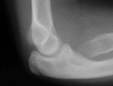 Lateral radiograph of elbow of patient A, depicting a dysplastic proximal radius. This is characteristic of dyschondrosteosis.
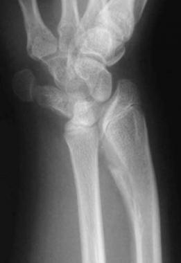 Preoperative anteroposterior radiograph of wrist of patient B. This patient has primary Madelung deformity (no sign of dyschondrosteosis).
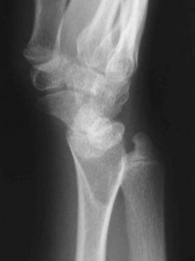 Preoperative lateral radiograph of wrist of patient B. The flame-shaped radiolucency in the metaphysis of the radius is occupied by the fibrocartilaginous Vickers ligament.
With such a similarity between primary MD and Leri-Weill dyschondrosteosis, the reason why many authors have described them as a single entity is apparent. However, many children with unilateral and bilateral MD have normal stature and no other characteristic of dyschondrosteosis. Therefore, it is reasonable to describe primary MD as a separate, but related, condition.
To differentiate primary MD from dyschondrosteosis, Felman and Kirkpatrick suggested the following criteria:[17] Primary MD can be defined as occurring in a child above the 25th percentile in height with no family history of dyschondrosteosis and no history of other secondary causes. Conversely, a patient who is shorter than 5 ft at skeletal maturity, has proximal involvement of the radius, and has a relatively short tibia and fibula may have mesomelic dwarfism.
Several hundred cases of MD have been presented in the literature since its first description. However, no published reports exist on the actual frequency of MD in the population.[18]
Goals for surgical correction of MD consist primarily of pain relief and correction of the cosmetic deformity. A secondary goal is to increase range of motion. Most patients who undergo biplane osteotomy with ulnar length adjustment or the crescentic radial osteotomy of Carter and Ezaki attain both primary goals.[19] Range of motion usually is only moderately improved at best. It should be noted that pain relief can be substantial and can be achieved quickly following release of the volar Vickers ligament.
Clinical Presentation
Copyright © www.orthopaedics.win Bone Health All Rights Reserved