Chondrosarcoma is a collective term for a group of tumors that consist predominantly of cartilage and that range from low-grade tumors with low metastatic potential to high-grade, aggressive tumors characterized by early metastasis. See the image below.
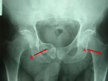 Plain radiograph shows low-grade chondrosarcoma in pelvis (B). Incidental finding is that proximal femur contains benign enchondroma (A).
Different types of chondrosarcoma have been described, as follows:
Benign cartilage lesions can be difficult to differentiate from slow-growing, low-grade chondrosarcomas. Secondary chondrosarcoma can occur in a previously benign cartilaginous lesion.
Chondrosarcomas can be classified into the following three histologic grades, depending on findings of cellularity, atypia, and pleomorphism:
The higher the grade, the more likely the tumor is to spread and metastasize. Grade I lesions rarely metastasize, whereas 10-15% of grade II lesions and more than 50% grade III lesions metastasize.
Clinical features of chondrosarcomas are as follows:
The workup rests primarily on diagnostic imaging modalities (eg, plain radiography, as well as CT and MRI).
Plain radiography
Secondary malignant degeneration should be suspected when sequential follow-up radiographs of benign cartilage tumors show the following findings:
MRI
CT scanning
Biopsy
Performing a truly representative biopsy of a chondrosarcoma is challenging because the lesion is composed of areas that carry different histologic grades. Identification of the most aggressive component of the tumor is critical. Considerations when performing biopsy are as follows:
The Enneking staging system for musculoskeletal sarcomas is applicable to chondrosarcomas, as follows[3] :
Surgery is the primary treatment for any chondrosarcoma. Complete, wide surgical excision of the chondrosarcoma is the preferred method when it is feasible. Radiotherapy and chemotherapy play limited roles in primary treatment. An exception is their use as adjuvant therapy or palliative treatment for tumors in surgically inaccessible areas or diffuse metastasis.
NextChondrosarcoma is a tumor of mesenchymal origin that predominantly is made of cartilage; it is the second most common primary malignant tumor of the bone. Chondrosarcoma is a collective term that encompasses a group of heterogeneous lesions with diverse morphologic features and clinical behaviors.[4] These lesions range from low-grade tumors with low metastatic potential to high-grade, aggressive tumors characterized by early metastasis.
The term chondrosarcoma should be used for a malignant tumor of the cartilage when the tumoral matrix is entirely cartilage. If the tumor exhibits bone-forming elements and primitive mesenchymal elements in addition to cartilaginous differentiation, it should not be classified as a chondrosarcoma, because its clinical behavior and therapeutic responses differ from those of a primary malignant chondrosarcoma.
Tumors with the aforementioned elements (ie, the presence of bone-forming elements and primitive mesenchymal elements in addition to cartilaginous differentiation) usually behave like chondroblastic osteosarcomas and are more aggressive than the conventional chondrosarcomas.
Different types of chondrosarcoma have been described, as follows:
Despite various investigations, it may be difficult to differentiate a benign cartilage lesion from a slow-growing, low-grade chondrosarcoma. Secondary chondrosarcoma can occur in a previously benign cartilaginous lesion.
Chondrosarcomas may be divided into primary and secondary lesions on the basis of their origins.[5] Primary chondrosarcomas arise de novo, whereas secondary chondrosarcomas arise from preexisting lesions of the cartilage.
Except for their origin in preexisting cartilaginous conditions, secondary chondrosarcomas are similar to conventional chondrosarcomas in all respects. In addition, the genes responsible for the lesions depend on the primary benign cartilaginous condition. Secondary chondrosarcomas occur in individuals with Ollier disease, Maffucci syndrome, multiple hereditary exostosis (diaphyseal aclasis), solitary osteochondroma, solitary enchondroma, solitary periosteal enchondroma, Paget disease, or radiation injury.
Bovee et al reported that most peripheral chondrosarcomas had a higher proliferation rate on Ki-67 immunohistochemistry and that they were associated with loss of heterozygosity at many loci.[6, 7] Only a few chondrosarcomas had anomalies, which were restricted to 9p21, 10, 13q14, and 17p13. These anomalies were peridiploid or near-haploid. Structural chromosomal aberrations and genetic instability were seen during cytogenetic analysis of well-differentiated, grade I chondrosarcomas. Nearly all grade III and some grade II chondrosarcomas were aneuploid.
Amplification of the c-myc proto-oncogene[8] and fos/jun[9] has also been implicated in the pathogenesis of chondrosarcoma.
With extraskeletal myxoid chondrosarcomas,[10] the t(9;22)(q22;q12) translocation is common, though t(9;17)(q22;q11.2) has also been described. Numerous genetic alterations have been found for dedifferentiated chondrosarcomas, but a shared loss of chromosome 13 suggests that the differentiated and dedifferentiated components originate from a common precursor.
Chondrosarcomas can be classified into the following three histologic grades, depending on findings of cellularity, atypia, and pleomorphism:
The higher the grade, the more likely the tumor is to spread and metastasize. Grade I lesions rarely metastasize, whereas 10-15% of grade II lesions and more than 50% grade III lesions metastasize.
Low-grade chondrosarcomas resemble benign cartilaginous tumors, and it is difficult to differentiate the two lesions on the basis of histologic features alone. The essential differences are the limited growth potential of benign cartilaginous tumors and the slow growth capacity of low-grade chondrosarcomas.
Dedifferentiated chondrosarcomas are more aggressive than grade III conventional chondrosarcomas.
Conventional central chondrosarcomas account for nearly 80-90% of all chondrosarcomas and 20-27% of all primary bone sarcomas[11] .They demonstrate a predilection for the axial skeleton. Rates of involvement are as follows:
The spine and the craniofacial bones are rarely involved.
Dedifferentiated chondrosarcomas are responsible for as many as 10% of all chondrosarcomas. The femur is the site most commonly involved, accounting for one-third of all dedifferentiated chondrosarcomas. The other sites of involvement are the pelvis (20%), the humerus (16%), the ribs (7%), and the scapula (7%).
Clear cell chondrosarcomas account for fewer than 5% of all chondrosarcomas. They have a predilection for the ends of long tubular bones, involving the epiphysis. Like chondroblastomas, these lesions extend to involve the articular cartilage. The proximal aspect of the femur is the site most often affected (45%), followed by the proximal portion of the humerus.
Fewer than 2% of all chondrosarcomas are mesenchymal chondrosarcomas. The maxilla and the mandible are the most common sites of involvement, followed by the vertebrae, the ribs, the pelvis, and the humerus. The appendicular skeleton is rarely involved.
Juxtacortical chondrosarcomas are rare and generally involve the surface of the diaphysis or metaphysis of long tubular bones.
Incidences do not differ among ethnic groups. Sex and age distributions are listed in Table 1 below.
Table 1. Sex Ratios and Ages of Peak Incidence for Different Types of Chondrosarcoma (Open Table in a new window)
Chondrosarcoma Male-to-Female Ratio Age of Peak Incidence Conventional Almost 1:1 (slight male predominance) 50-70 y (most common > 50 y, gradual increase with age) Dedifferentiated Similar to the ratio above > 50 y Clear cell 2.4:1 20-40 y (common 10-90 y) Mesenchymal 1:1 20-30 y (common in teenagers and young adults) Juxtacortical 1:1 20-40 yThe prognosis is correlated with the grade and stage of the lesion at the time of diagnosis.[12] The location of the lesion is also important because tumors in areas where complete wide resection is possible are associated with better prognoses. In general, chondrosarcomas of the head and neck are associated with better disease-specific survival and overall survival rates than chondrosarcomas located elsewhere.[13]
Recurrence and distant metastasis may develop. The metastasis rate for primary chondrosarcoma is higher than that for secondary chondrosarcoma, and the rate of distant metastasis is higher in patients with local recurrence than in those without local recurrence.
Mortality and morbidity data for the various types of chondrosarcomas are summarized below.
Evans et al[14] showed that the survival rate depends on the histologic grade of the tumor, as follows:
Overall, the 5-year survival rate for conventional chondrosarcomas is 48-60%.[11] Intralesional surgery is not advised even in grade I lesions, especially in the pelvis, because the local recurrence rate is 100% in such cases.
Grade I tumors do not metastasize, whereas 66% of grade III tumors do. The most common sites for metastases are the lungs. Recurrences typically appear 5-10 years or longer after surgery.
Dedifferentiated chondrosarcoma is highly lethal. It is associated with a 10% survival rate after 1 year. Even with early surgical treatment, disseminated hematogenous metastasis occurs in most patients.[11]
Although clear cell chondrosarcomas are low-grade tumors, they can lead to distant metastasis. Late recurrences (>10 years) have been described. Overall, the recurrence rate is 16%[11] .
The 5-year survival rate for mesenchymal chondrosarcoma is less than 50%, with an overall 10-year survival rate of 28%.[11]
Juxtacortical chondrosarcomas are low- to intermediate-grade lesions; the prognosis for these tumors is better than that for other tumors.[11]
Clinical Presentation
Copyright © www.orthopaedics.win Bone Health All Rights Reserved