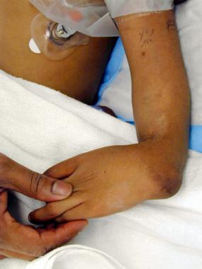
Radial clubhand is a deficiency along the preaxial or radial side of the extremity. Although considerable forearm and hand anomalies are the classic findings, proximal deficiencies also can occur throughout the arm and shoulder girdle. The elbow abnormalities can include deficiences of the olecranon, capitellum, coronoid fossa, and medial epicondyle.
In 1733, Petit first described radial clubhand in an autopsy of a neonate with bilateral clubhands and absent radii. Subsequent autopsy observations detailed the anomalous anatomy associated with radial clubhand and the associated malformations of other body systems.
Initial surgical treatment of radial clubhand involved an ulnar osteotomy to correct the bow, along with splitting of the distal ulna for insertion of the carpus. Reconstruction of the radius with a bone graft to support the carpus was reported in the 1920s, and nonvascularized epiphyseal transfer was reported in 1945. Results of these procedures were disappointing. They had multiple causes of failure, including disruption of the ulnar growth plate and subsequent increase in limb-length discrepancy, inadvertent ankylosis or arthrodesis of the wrist and loss of motion, and failure of the transplanted bone to grow, with eventual loss of radial support.
Centralization of the carpus on the distal ulna has emerged as the preferred surgical technique for correcting radial clubhand. Pioneers in congenital hand surgery developed the basis for this procedure. Numerous modifications have been described to obtain or maintain correction of the wrist on the ulna.
Radialization is a technique that involves overcorrection of the carpus on the ulna combined with tendon transfer to further rebalance the wrist.[1]
Despite over 250 years of investigation, current treatment regimens are still unable to restore normality: The limb remains short, and the wrist lacks full function.
This article summarizes the history of radial deficiencies, lists potential etiologies, highlights relevant pathoanatomy, discusses treatment regimens, reviews expected outcome, and details potential complications.
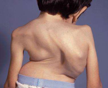
NextClinical features of radial deficiency (types II, III, and IV; see Presentation, Classification) are dramatic, with abnormalities of the entire extremity. The scapula is often small, and the clavicle is often shorter, with an increased curvature. The humerus may or may not be short, and deficiencies of the capitellum and trochlea are common. Elbow motion is usually diminished more in flexion than in extension.
The forearm is always decreased in length, and the ulna is approximately 60% of the normal length at the time of birth. This discrepancy persists throughout the growth period and into adulthood. True forearm rotation is absent in patients with partial or complete aplasia of the radius.
The wrist is radially deviated and develops a perpendicular relation to the forearm over time. The articulation between the carpus and ulna is usually fibrous and abnormal, though some hyaline cartilage can be found. Wrist motion is primarily in the radial/ulnar plane, with some flexion/extension. Ossification of the carpal bones is delayed, with the scaphoid and trapezium often absent or hypoplastic. The capitate, hamate, and triquetrum are usually present but ossify late.
The fingers are often stiff, with limited motion at the metacarpophalangeal (MCP) and interphalangeal (IP) joints. The preaxial index and long fingers are more affected than the postaxial ring and small digits.
Numerous muscular abnormalities are found throughout the upper extremity. The deltoid or the pectoralis major can be hypoplastic, can be partially absent, or can have an abnormal insertion. The biceps may be absent or fused to the underlying brachialis.
The forearm demonstrates the most severe abnormalities, which may involve any of the muscles that originate from or attach to the radius, including the following:
The extrinsic flexors and extensors of the fingers are usually adherent, with abnormal origins and insertions. The flexor and extensor carpi ulnaris, as well as the interossei, lumbricals, and hypothenar muscles, are often normal, whereas abnormalities of the thumb muscles are more related to the degree of thumb hypoplasia.
The radial nerve usually terminates at the elbow, and the ulnar nerve is normal. An enlarged median nerve substitutes for the absence of the radial nerve and supplies a dorsal branch for dorsoradial sensibility. This subcutaneous branch is positioned in the fold between the wrist and forearm and must be protected during surgery. The vascular anatomy demonstrates a normal brachial and ulnar artery. The radial artery is often absent, and the interosseous arteries usually remain patent.
Radial deficiency is associated with numerous systemic conditions,[2] including Holt-Oram syndrome (cardiac septal defects); TAR syndrome; Fanconi anemia (aplastic anemia); and VACTERL syndrome.[3, 4] In addition to these conditions, a variety of associated musculoskeletal deformities appear sporadically. These include cleft palate, clubfoot, kyphosis, scoliosis, torticollis, and rib deformities.
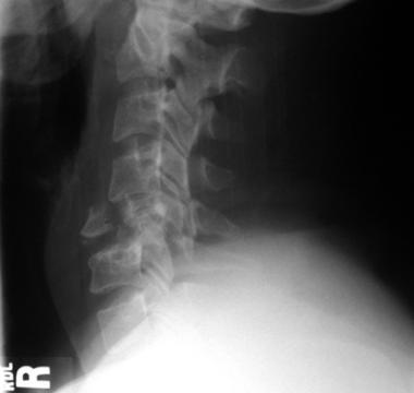
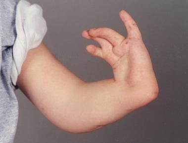
The forearm is foreshortened, and the wrist is positioned in radial deviation. This malalignment assumes a perpendicular relation over time (see the image below).
 Perpendicular relationship between wrist and forearm in radial clubhand.
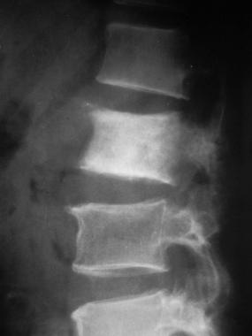
The right-angle position (see the image below) further shortens the limb and limits the ability to reach into space. The awkward angulation between the wrist and forearm places the extrinsic flexors and extensors at a mechanical disadvantage. The tendons must traverse this angle to elicit finger motion, and this limits the ability to move the digits.
 Right angle between wrist and forearm.
In the 19th century, the etiology of radial clubhand was theorized to be either a congenital absence or an acquired defect secondary to syphilis. In 1895, Kummel proposed the cause to be abnormal pressure upon the embryo along the radial bud between the third and seventh week of gestation.
Current theory relates the etiology of radial clubhand to the apical ectodermal ridge (AER). This structure is a thickened layer of ectoderm that directs differentiation of the underlying mesenchymal tissue and limb formation. Removal of a portion of the AER in chick embryos has produced anomalies similar to radial clubhand.[5] Therefore, a defect of the AER is the most probable cause of radial clubhand, with the extent of deformity related to the degree and extent of the AER absence.[6]
The reported incidence of radial clubhand ranges from 1 in 55,000 to 1 in 100,000 live births. Most cases are sporadic without any definable cause. However, exposure to teratogens, such as thalidomide and radiation, can yield radial deficiencies.
Radial deficiency is bilateral in 50% of cases and is slightly more common in males than in females (3:2). The incidence of radial deficiency within the same family is low, ranging from approximately 5% to 10% of reported cases; it is most common in radial aplasia associated with cardiac abnormalities.
Comparison of results following centralization is difficult because of the many surgical modifications, variations in technique to determine the degree of deformity, and potential inconsistencies in measurement. Measurements to determine the deformity have been discussed by numerous authors.[7, 8]
Heikel described the construction of lines to calculate ulna curvature and overall deformity.[9] He also discussed the difficulties with measurements of angles in radial clubhand, including inexact determination of lines and the difficulties in obtaining a standardized radiographic view of the radial clubhand.
Manske et al, as well as Bayne and Klug, recommended the hand-forearm angle using the longitudinal axis of the third metacarpal and distal ulna, whereas Bora et al used the ulnar bow to assess deformity and recurrence.[10, 11, 12, 13] Damore et al used total angulation, which is the combination of radial deviation of the hand and the ulna bow.[14] This represents the clinical deformity by combining the radial deviation of the wrist and ulna bow.
Lamb monitored 31 centralizations for an average of 5 years and measured a preoperative radial deviation of 78° and a follow-up angle of 22°.[15] Bora et al reported on 14 extremities at an average of 14.6 years after centralization and showed a gradual increase in the hand-forearm angle to an average of 25°.[13] Manske et al reported on 21 radial clubhands and measured the hand-forearm angle at 58° preoperatively and 26° at an average 34-month postoperative period.[10]
Watson et al monitored 12 centralizations for 10 years and reported a recurrence to an average of 30°.[16] Bayne and Klug monitored 53 patients for an average of 8.6 years and reported 81% good or satisfactory results, defined as a hand-forearm angle of less than 30°.[11] Damore et al monitored 14 patients for an average of 7 years.[14] An initial significant improvement from a preoperative total angulation of 83° to an immediate postoperative angle of 25° was noted. At follow-up, total angulation had increased to 63°, with a 38° loss of correction.
Kotwal et al, in a retrospective analysis of 446 patients with type III and IV radial longitudinal deficiency treated over a 20-year period, found that patients treated with surgery had better appearance, function and performed better with activities when compared to those treated with stretching and splinting alone.[17]
In Vilkki’s long-term follow-up of 19 wrists followed for over 11 years treated with vascularized second metatarsophalangeal transfer, average final hand-forearm angle was 28º of radial deviation.[18] Total active wrist motion averaged 83º (range, 30-115º). Overall ulnar growth averaged 15.4 cm, and overall relative ulnar length was 67% of the contralateral side (range, 51-78%).
Clinical Presentation
Copyright © www.orthopaedics.win Bone Health All Rights Reserved