Phalangeal fractures are common injuries that may significantly affect hand function.[1] Even subtle injuries, such as a simple finger jam, can lead to decreased motion and a poor outcome if not diagnosed and treated promptly. This is especially true with injuries to the proximal interphalangeal joint (PIPJ). Fractures of the phalanges, if unstable, necessitate fixation secure enough to allow early motion in order to prevent adhesion formation.
Historically, closed treatment has been the therapeutic mainstay. Percutaneous pinning allowed the conversion of more unstable fracture patterns to stable configurations capable of tolerating early motion. Subsequently, minifragment screws and plates were developed to assist in the management of complex phalangeal fractures.
For patient education resources, see the First Aid and Injuries Center, as well as Broken Finger and Broken Hand.
NextFew places in the body exist where function and anatomy are as closely intertwined as they are in the finger. Injuries and subsequent scar formation can upset the delicate balance that normally exists, particularly at the PIPJ and the extensor apparatus. Anatomic considerations are based on the level of injury.
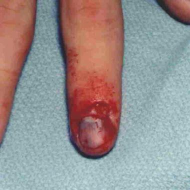
The terminal extensions of the flexor and extensor tendons insert into the base of the distal phalanx. These tendons can rupture at their insertion or can avulse a fragment of bone. Tuft fractures are commonly associated with injury to the overlying nail bed.
The head of the middle phalanx consists of two condyles that articulate with the base of the distal phalanx. With an axial load, one or both of the condyles may fracture. A closely adherent volar plate provides significant stability. Radial and ulnar collateral ligaments provide resistance to stresses in the coronal plane.
Sublimis tendons insert along a broad expanse on the volar aspect of the proximal half of the phalanx. The profundus tendon is held tightly in the flexor sheath by the important A4 pulley at the midportion of the phalanx. The middle phalanx region also contains additional cruciate pulleys (C2 and C3), which are located proximal and distal to the A4 pulley, respectively.
On the extensor side, the central slip inserts into the base of the middle phalanx. The lateral bands join over the distal portion of this phalanx to form the terminal extensor tendon. The two lateral bands are held together by the triangular ligament, which prevents volar subluxation of the lateral bands.
The anatomy of the PIPJ is similar to that of the distal interphalangeal joint (DIPJ). The volar plate covers a broad expanse over the joint and is the main stabilizer to joint dislocation. The collateral ligaments are larger at the PIPJ and consist of proper and accessory components.
Sublimis and profundus tendons run together in the flexor sheath (zone 2) at this level. The A2 flexor pulley covers most of the proximal half of the phalanx; the C1 pulley is located more distally. The extensor digitorum communis tendon runs the length of the phalanx and is stabilized by oblique and transverse fibers of the intrinsic apparatus. The lateral bands run from a lateral and volar position at the proximal aspect of the phalanx to a more dorsolateral position at the level of the PIPJ.
The stability of phalangeal fractures is dependent on the following factors:
Distal tuft fractures usually are stable, despite comminution. Unicondylar and bicondylar fractures involving the interphalangeal joints are inherently unstable. Displaced fractures involving the diaphyses of the proximal and middle phalanges also are unstable secondary to the pull of the intrinsics and flexor tendons. Fractures with an intact periosteal sleeve and no initial displacement usually are stable.
Fractures and dislocations of the phalanges occur from a variety of mechanisms. In younger patients, these injuries are more likely to be sports related. Older patients are likely to be injured by machinery or by falls. Crush injuries are common at the distal phalanx, whereas the PIPJ is usually damaged by an axial blow to the finger.[2]
Because many injuries to the phalanges go unreported, defining a true incidence is difficult. Fractures of the phalanges certainly are among the most common in the entire skeleton and may account for as many as 10% of all fractures.[2]
Outcome following phalangeal fractures depends on patient and injury factors, as well as on the expertise of the surgeon. Poorer results have been documented for patients older than 50 years and for those with associated systemic illness. High-energy fractures with comminution and soft tissue injury also lead to poorer outcomes. Tendon injury, especially extensor tendon injury, in association with fracture, compromises results. Factors that the surgeon can control include selecting the appropriate fixation and ensuring that the period of immobilization does not exceed 3 weeks.
Clinical Presentation
Copyright © www.orthopaedics.win Bone Health All Rights Reserved