Disorders of the peroneal tendons are often overlooked as causes of lateral ankle pain and have been reported infrequently. The injuries may go unrecognized or may be misdiagnosed as ankle sprain. The three major pathologies of the peroneal tendons include the following:
Monteggia described peroneal tendon subluxation in 1803,[1] and this entity seems to be more commonly encountered than are disruptions of the peroneus longus or brevis alone. Nonetheless, peroneus brevis disorders have been described more often in the literature, with peroneus longus problems gaining more recent attention. However, much of the literature regarding both tendons is in the form of case reports.
For patient education resources, see Ankle Sprain.
NextThe peroneal muscles make up the lateral compartment of the leg and are innervated by the superficial peroneal nerve. The peroneus longus originates from the lateral condyle of the tibia and the head of the fibula. The tendon of the peroneus longus courses behind the peroneus brevis tendon at the level of the ankle joint, travels inferior to the peroneal tubercle, turns sharply in a medial direction at the cuboid bone, and passes inferior to it in the cuboid tunnel, inserting into the lateral aspect of the plantar first metatarsal and the medial cuneiform. (See the image below.)
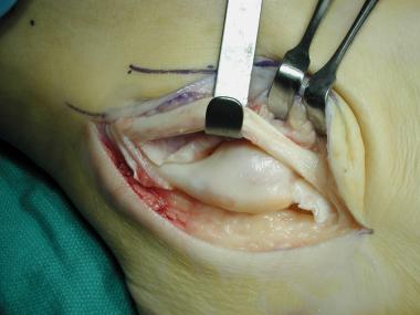 Peroneus longus tendon next to peroneus brevis tendon.
A sesamoid bone called the os peroneum may be present within the peroneus longus tendon at about the level of the calcaneocuboid joint. The frequency with which an os peroneum occurs is controversial, with many supporting the idea that one is always present. However, the os peroneum may be ossified in only 20% of the population. The peroneus longus serves to plantarflex the first ray, evert the foot, and plantarflex the ankle.
The peroneus brevis originates from the fibula in the middle third of the leg. Its tendon courses anterior to the peroneus longus tendon at the ankle. It courses over the peroneal tubercle and inserts onto the base of the fifth metatarsal. The peroneus brevis everts and plantarflexes the foot.
The peroneal tendons share a common tendon sheath proximal to the distal tip of the fibula. More distally, each tendon is housed within its own sheath. The common sheath is contained within a sulcus on the posterolateral aspect of the fibula, which prevents subluxation.
The retromalleolar sulcus/groove is 6-7 mm wide and 2-4 mm deep and is lined by fibrocartilage to allow smooth gliding of the tendons. The sulcus is concave in 82% of cases, flat in 11%, and convex in 7%. A rim of fibrocartilage is often present at the lateral border of fibula, adding 2-4 mm to the depth of the groove.
At the level of the retromalleolar sulcus, the superior peroneal retinaculum (SPR) and the calcaneofibular ligament stabilize the peroneal tendons. Therefore, inversion injuries that damage the calcaneofibular ligament can also result in injury to the SPR. More distally (inferiorly) the tendons are restrained by the inferior peroneal retinaculum (IPR).
The SPR is the primary restraint to tendon subluxation. This fibrous band originates on the posterolateral aspect of the fibula and inserts onto the lateral wall of the calcaneus. It is reported to average 10-20 mm in width and to course in a posteroinferior direction, though variants are not uncommon.
Problems may arise in either of the peroneal tendons alone, or both may be involved with subluxation. The hallmark of disorders of the peroneal tendons is laterally based ankle or foot pain. Whether the problem is tendinous degeneration or subluxation, the clinical manifestation is pain. With time, loss of eversion strength may occur.
Problems arising with the peroneus longus include tenosynovitis and tendinous disruption (acute or chronic). The os peroneum may be involved with the degenerative process or as a singular disorder and can be fractured or fragmented. Longitudinal tears of the peroneus longus are uncommon but have been reported.[2]
Longitudinal tears of the tendon are the most common problem seen with the peroneus brevis tendon. These may be single or multiple. Tendinitis and tenosynovitis also may occur.
Subluxation of both peroneal tendons may occur after an acute traumatic episode or may be of a more chronic nature.
Brandes and Smith described and classified primary peroneus longus tendinopathy,[3] specifying the following three anatomic zones in which the tendon can be injured:
In their series, complete ruptures were most likely in zone C, whereas partial ruptures were more common in zone B.[3] In the same study, surgical findings were classified into three groups, as follows:
Other attempts have been made to classify peroneal tendon pathology. Sobel et al presented a classification for tears of the peroneus brevis tendon that divides these injuries into the following four grades[4, 5] :
However, this grading system has no implications for clinical management.
Eckert and Davis classified SPR pathology as follows[6] :
The precise etiology of peroneal tendon disorders depends somewhat on the specific problem being addressed. All disorders may result following a traumatic episode, direct or indirect, with a lateral ankle sprain being the most common trauma. Severe calcaneal fractures with lateral displacement of the calcaneus can also result in the dislocation of the peroneal tendons. Forceful dorsiflexion of the foot with the peroneal muscles strongly contracted, as happens with a forward fall during skiing or springboard diving, can result in acute subluxation or dislocation of the peroneal tendons.
Brandes and Smith reported that 82% of patients with primary peroneus longus tendinopathy had a cavovarus hindfoot.[3] The presence of an os peroneum also has been postulated to predispose to peroneus longus rupture. Ruptures likewise have been reported to occur secondary to rheumatoid arthritis and psoriasis, as well as diabetic neuropathy, hyperparathyroidism, hypothyroidism, foot and ankle fractures, tophaceous gout, fluoroquinolone use, and local steroid injection.[7, 8, 9]
A hypertrophied peroneal tubercle that increses mechanical stresses on the peroneal tendons and interferes with normal gliding within the tendon sheaths can also predispose to peroneal tendon attrition and rupture. Irregularities of the retromalleolar groove can result in peroneal tendon tears as well.
Longitudinal splits in the peroneus brevis tendon appear to result from mechanical factors. Repetitive or acute trauma causes the attritional ruptures. These ruptures may result from an incompetent SPR that allows the peroneus brevis to rub abnormally against the fibula.
Overcrowding from a peroneus quartus muscle, peroneal tenosynovitis, or a low-lying muscle belly of the peroneus brevis also has been reported as a cause of tendon ruptures. The blood supply to the tendon has been controversial, with cases for both tenuous and adequate blood supply having been put forth by various authors; at present, the issue remains undecided.
Tenosynovitis and tendinitis of the peroneal tendons are common among runners and ballet dancers and may occur in more than 75% of patients with chronic lateral ankle instability.
Subluxation of the peroneal tendons results from disruption of the SPR and usually involves avulsion of the retinaculum from its fibular insertion. The mechanism of injury typically involves an inversion injury to the dorsiflexed ankle with concomitant forceful contraction of the peroneals. Some patients have a more chronic presentation and cannot recall a traumatic episode. Congenital dislocations also have been reported. An inadequate groove for the peroneals in the posterolateral fibula may be a cause of subluxation as well.
Pathology of the longus and brevis tendons almost always occurs concurrently. Brandes and Smith noted a 33% incidence of peroneus longus tendon tears in cases with peroneus brevis tendon tears.[3]
Disorders of the peroneal tendons are less common than other tendon problems involving the Achilles or posterior tibial tendons. However, it is impossible to estimate their true frequency in the United States or abroad.
Outcome after surgical treatment of peroneal tendon pathology is difficult to accurately assess. Much of the literature is in the form of case reports, with few large series in existence. Additionally, with the large variability in treatment, conclusions are hard to draw. Much of the decision-making in this area is based on surgeon experience.
Sammarco reported 10 of 13 patients with good or excellent results using varied surgical treatments of peroneus longus problems.[10] Eight of those patients also had associated peroneus brevis pathology. Thompson and Patterson reported on three of their patients with peroneus longus pathology who responded well to surgical debridement and tenodesis.[11]
Krause and Brodsky reviewed 20 cases of peroneus brevis tears and noted that good or excellent results can be expected with surgical treatment.[12]
Demetracopoulos et al conducted a review of long-term outcomes in 34 patients with peroneal tendon tears treated with debridement and primary repair.[13] At the time of final follow-up, pain scores had improved from a mean of 39 before operation to a mean of 10 postoperatively. There was a significant increase in the Lower Extremity Functional Scale score, from a mean of 45 preoperatively to a mean of 71 postoperatively. Of the 18 patients who responded, 17 returned to full sporting activity without limitation. There were no reoperations or operative failures.
Current data confirm good outcomes, high satisfaction, and a quick return to sports with surgical treatment of peroneal tendon dislocation. Rates of return to sports are significantly higher in patients treated with both groove deepening and SPR repair than in those treated with SPR repair alone.[14] Whereas a redislocation rate of nearly 50% is reported with conservative treatment, the rate is lower than 1.5% at long-term follow-up after surgical treatment.
Clinical Presentation
Copyright © www.orthopaedics.win Bone Health All Rights Reserved